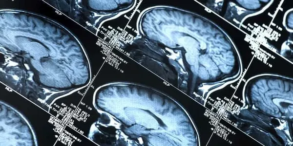
Research has shown us that it is the diseases themselves that cause much of the cognitive dysfunction. For many years, people thought that cognitive problems were secondary to other symptoms, such as psychosis, lack of motivation or unstable mood, but now we know that this is not the case.
Cognitive dysfunction is a major symptom of schizophrenia and some mood disorders. This is why cognitive problems are evident even when other symptoms are controlled for, even when people are not psychotic or in a mood episode.
In addition, research has shown that parts of the brain used for specific cognitive skills often do not function normally in people with schizophrenia and certain affective disorders. This indicates that mental illness affects the way the brain works and that is what causes cognitive problems. There are many myths about mental illness and cognitive dysfunction. Some of the most common ones are listed in the sidebar below.
The ability to pay attention, remember, and think clearly is ultimately the result of a complex interplay of factors. While it is true that mental illness often causes cognitive impairment, it is also true that other factors will affect thinking skills. Most people think better, pay attention, and remember better when they are not emotionally stressed and when they have had the opportunity to learn adaptive cognitive skills.

Among the comorbidities associated with epilepsy, cognitive abnormalities are among the most common and problematic.
In people with epilepsy there is a high associated rate of cognitive difficulties that compromise educational progress and achievements throughout life. In addition to a higher incidence of low IQ, in approximately half of children with epilepsy there is an identified discrepancy between IQ and performance. Children who have poorly controlled (drug-resistant) seizures are more likely to have lower IQ scores than children with well-controlled seizures. Adults with chronic epilepsy are also vulnerable to cognitive regression.
Both children and adults with epilepsy frequently complain of memory disorders.
People with epilepsy may have transient epileptic amnesia, in which the sole or main feature of the epilepsy is episodic amnesia, accelerated long-term forgetting, in which newly acquired memories fade over days or weeks, and long-term memory impairment, in which public or autobiographical events are forgotten.
Accelerated long-term forgetting is a condition in which individuals learn and initially retain information normally, but forget the information at an unusually fast rate. Both accelerated forgetting and long-term memory impairment are primarily seen in people with temporal lobe epilepsy.

“What’s good for your heart is good for your brain.” Evidence supports preventing or controlling cardiovascular conditions such as high blood pressure to protect brain health as adults age.
One in three adults has high blood pressure, putting them at risk for heart disease and stroke — conditions that are among the leading causes of death. High blood pressure (also called hypertension) can also significantly affect brain health. That's reason enough to monitor your blood pressure regularly and treat it if it's high.
Observational studies show that having high blood pressure in middle age (ages 40 to 60) increases the risk of cognitive decline later in life.
The blood flow that keeps the brain healthy can, if reduced or blocked, damage this essential organ. Uncontrolled high blood pressure plays a role in this damage. Over time, the force of blood pushing through the arteries can cause blood vessels to become scarred, narrowed, and diseased. This damage can impede blood flow to many parts of the body, including the brain.
The types of pathologies that high blood pressure leads to include cerebrovascular damage, such as a major stroke, a series of small strokes, reduction of white and gray matter and microinfarcts (tiny areas of dead brain tissue), and possibly the plaques and tangles typical of Alzheimer's.

Diabetes has been known to have effects on the brain for over a hundred years. In the early 20th century, researchers and physicians recognized that people with diabetes frequently complained of memory and attention problems. In 1922, it was shown that people with diabetes performed poorly on cognitive tasks that tested memory and attention. The term “diabetic encephalopathy” was introduced in 1950 to describe complications of diabetes related to the central nervous system.
Other terms such as functional brain impairment and central neuropathy have also been used in the literature to describe diabetes-related cognitive dysfunction.

Sleep apnea syndrome is a breathing disorder during sleep, characterized by total or partial obstruction of the upper airway leading to hypoxia or hypercapnia, in addition to an increase in respiratory effort. These features produce microarousals that result in sleep disruption and changes in neuronal activity. All of these are potential mechanisms for cognitive impairment.
Nocturnal symptoms include loud snoring, unrefreshing sleep, nocturia, sweating, and dry mouth. One of the most common daytime symptoms in patients with OSA (obstructive sleep apnea) is daytime sleepiness. This greatly influences quality of life and cognitive performance.
OSA has been associated with a wide range of psychological problems such as depression, anxiety, neurocognitive dysfunction, especially attention, alertness, memory and learning, phenomena due to sleep fragmentation and intermittent hypoxemia. Sleep fragmentation, sleep deprivation and the association of excessive daytime sleepiness are proposed mechanisms underlying cognitive impairment through their impact on attention.
The exact prevalence of cognitive disorders and their severity are unknown due to the multiple comorbidities with which this syndrome is associated in adult patients with OSA.
Studies on the cardiovascular effects of OSA have shown that the disorder produces changes in vascular structure and function, changes that are frequently found in other hypoxic populations. It is assumed that hypoxia would have a direct effect on the neuropsychic in patients with OSA, with similar mechanisms existing in terms of cardiovascular changes and cerebral vessels. Hypoxia produces immediate vasodilation, being a protective mechanism to more efficiently distribute oxygen to the affected organ. Studies have shown that this protective mechanism does not exist in patients with OSA. One possible reason why these patients do not respond to hypoxemia is because they suffer repeated episodes (more than five events/h) and desaturation, not just a sustained hypoxic event. Therefore, since the recovery time after the episode is limited, it is not possible to estimate whether there is a protective response to recurrent hypoxic events, but it is assumed that the vessels would suffer.
Therefore, in patients with OSA, there are hypoxic and reperfusion injuries with increased lipid peroxidation. This process involves the oxidation of polyunsaturated fatty acids with the formation of reactive oxygen species and toxic products, which have potentially damaging effects on the brain and heart.
Furthermore, in patients with vascular pathology, endothelial dysfunction is present. In patients with OSA, imbalances appear between vasoconstrictor mediators (higher levels of thromboxane and endothelin) and vasodilators (nitric oxide, prostacyclin) and nitric oxide production has been shown to decrease in OSA. This imbalance predisposes to atherosclerosis.
Therefore, the effects on cerebral blood flow, as well as hypoxia, can lead to the appearance of cerebral infarcts, resulting in vascular dementia. The presence of endothelial dysfunction, with the appearance of neurocognitive deficits, has even been described in studies carried out in the population.

There are ten cardiovascular disease risk factors that can affect the brain, each through distinct but overlapping vascular or cellular mechanisms, highlighting the broad impact that cardiovascular disease risk can have on brain structure and function. In terms of risk, blood pressure is perhaps the most studied factor. Associations between high blood pressure and poor performance on neuropsychological tests are well described, particularly in the areas of memory, attention, and executive function, a domain that involves higher-order cognitive processes such as reasoning, planning, cognitive flexibility, and initiation of appropriate cognitive processes.
Furthermore, in patients with vascular pathology, endothelial dysfunction is present. In patients with OSA, imbalances appear between vasoconstrictor mediators (higher levels of thromboxane and endothelin) and vasodilators (nitric oxide, prostacyclin) and nitric oxide production has been shown to decrease in OSA. This imbalance predisposes to atherosclerosis.
Therefore, the effects on cerebral blood flow, as well as hypoxia, can lead to the appearance of cerebral infarcts, resulting in vascular dementia. The presence of endothelial dysfunction, with the appearance of neurocognitive deficits, has even been described in studies carried out in the population.

Cancer-related cognitive changes and impairment may be due to the cancer itself. Impairments associated with brain tumors are often specific to the location of the lesion, such as occipital tumors resulting in visual impairments. The location and momentum of the lesion (i.e., the rate of tumor growth that may result in destruction, crowding, displacement, and infiltration of brain tissue) influence the presence, intensity, and pattern of the resulting cognitive changes in patients with brain tumors. Patients with high-grade gliomas often show greater overall cognitive impairment compared to those with low-grade gliomas, which could be attributed to greater invasion and/or increased pressure on nearby normal brain tissue.
Up to 90 percent of patients with brain metastases have some cognitive impairment before treatment, and the degree of impairment correlates with total lesion volume rather than the number of metastatic lesions.